Home

Store shuffle image mitochondria under light microscope cough Mammoth pivot

3D Label-free Live Cell Imaging: Mitochondria in Holotomography vs Electron  Microscopy - YouTube
3D Label-free Live Cell Imaging: Mitochondria in Holotomography vs Electron Microscopy - YouTube

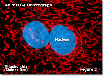
Molecular Expressions Cell Biology: Mitochondria

Electron microscopy morphology of the mitochondrial network in gliomas and  their vascular microenvironment - ScienceDirect
Electron microscopy morphology of the mitochondrial network in gliomas and their vascular microenvironment - ScienceDirect
Pin on Make: mitochondria

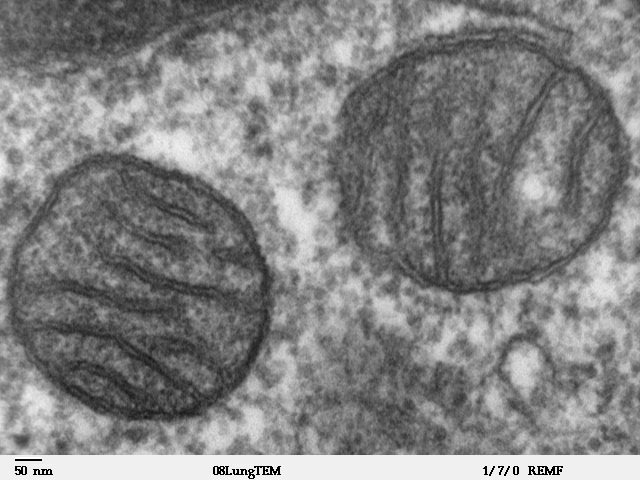
Mitochondrion - Wikipedia

Cell Micrographs | BioNinja

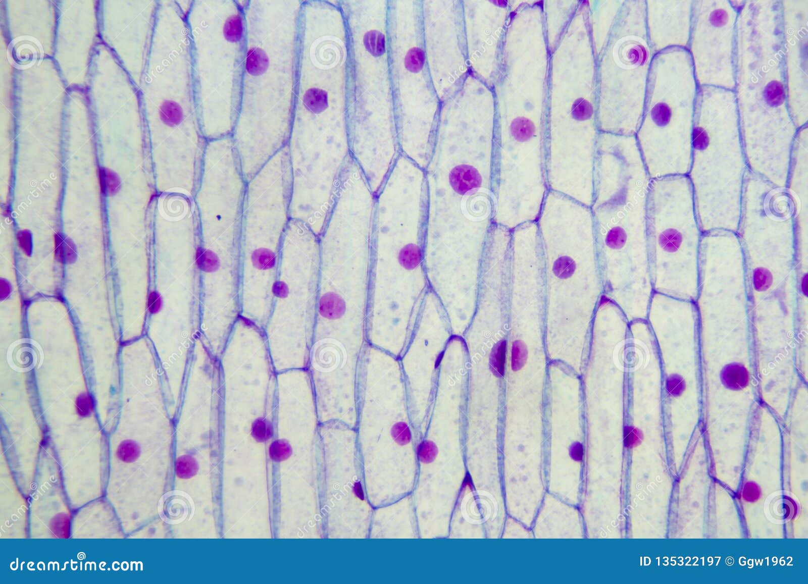
Plant mitochondria stock image. Image of experiment - 135322197

Insight into mitochondrial structure and function from electron tomography  - ScienceDirect
Insight into mitochondrial structure and function from electron tomography - ScienceDirect

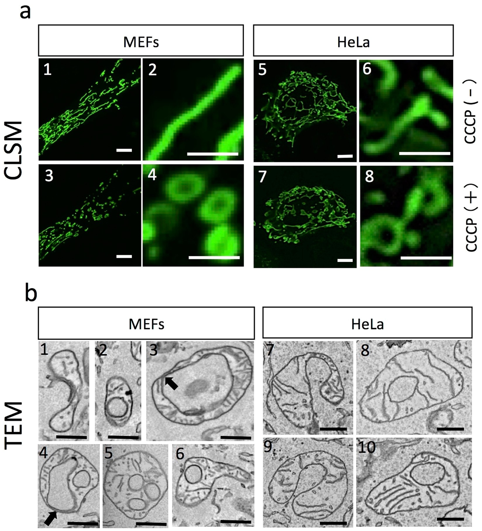
Uncoupled mitochondria quickly shorten along their long axis to form  indented spheroids, instead of rings, in a fission-independent manner |  Scientific Reports
Uncoupled mitochondria quickly shorten along their long axis to form indented spheroids, instead of rings, in a fission-independent manner | Scientific Reports

What cell organelles can be seen under the electron microscope but not with  the light microscope and their functions in the cell? - Quora
What cell organelles can be seen under the electron microscope but not with the light microscope and their functions in the cell? - Quora

Why can't we see cell organelles such as mitochondria, ribosomes, plastics,  etc under a compound microscope, although it is stained darker than  cytoplasm? - Quora
Why can't we see cell organelles such as mitochondria, ribosomes, plastics, etc under a compound microscope, although it is stained darker than cytoplasm? - Quora

Light and electron microscopy showing ultrastructural changes in the... |  Download Scientific Diagram
Light and electron microscopy showing ultrastructural changes in the... | Download Scientific Diagram

Light and electron microscopy of mitochondria in the oocytes of Argulus...  | Download Scientific Diagram
Light and electron microscopy of mitochondria in the oocytes of Argulus... | Download Scientific Diagram

Electron microscopy reveals cristae remodeling within mitochondria of... |  Download Scientific Diagram
Electron microscopy reveals cristae remodeling within mitochondria of... | Download Scientific Diagram

The morphology of mitochondria. (a) Thin-section electron micrograph of...  | Download Scientific Diagram
The morphology of mitochondria. (a) Thin-section electron micrograph of... | Download Scientific Diagram
A new glow for electron microscopy | MIT News | Massachusetts Institute of  Technology
A new glow for electron microscopy | MIT News | Massachusetts Institute of Technology

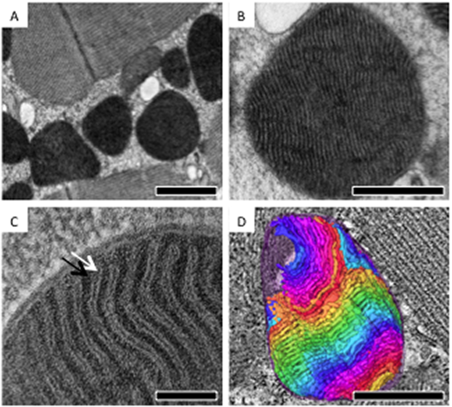
Electron tomographic analysis reveals ultrastructural features of  mitochondrial cristae architecture which reflect energetic state and aging  | Scientific Reports
Electron tomographic analysis reveals ultrastructural features of mitochondrial cristae architecture which reflect energetic state and aging | Scientific Reports

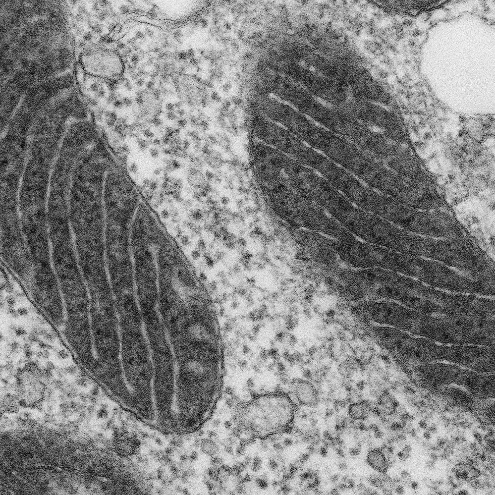
Tem mitochondria hi-res stock photography and images - Alamy

When we see a cheek cell through a microscope, can we see the mitochondria?  - Quora
When we see a cheek cell through a microscope, can we see the mitochondria? - Quora

4,188 Mitochondria Images, Stock Photos & Vectors | Shutterstock

Mitochondria: A worthwhile object for ultrastructural qualitative  characterization and quantification of cells at physiological and  pathophysiological states using conventional transmission electron  microscopy - ScienceDirect
Mitochondria: A worthwhile object for ultrastructural qualitative characterization and quantification of cells at physiological and pathophysiological states using conventional transmission electron microscopy - ScienceDirect

The Shrinking Mitochondrion | The Scientist Magazine®

Mitochondria Under Electron Microscope - YouTube

A mitochondria as seen with an electron microscope. Because you look at  small stuff with that tool, not at ants. : r/Damnthatsinteresting
A mitochondria as seen with an electron microscope. Because you look at small stuff with that tool, not at ants. : r/Damnthatsinteresting

Are mitochondria the natural cellular light sources?

Mitochondrion, SEM - Stock Image - C001/2592 - Science Photo Library

Light microscopic methods to visualize mitochondria on tissue sections -  ScienceDirect
Light microscopic methods to visualize mitochondria on tissue sections - ScienceDirect